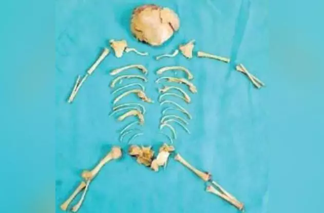
Vizag: KGH doctors perform rare surgery to remove ‘stone baby’ from woman’s womb
The woman reportedly became pregnant three years ago but attempted to terminate the pregnancy through an abortion
By Newsmeter Network
Visakhapatnam: Doctors at King George Hospital (KGH) in Visakhapatnam removed the skeleton of a fetus, known as a ‘stone baby’ or `lithopedion’ from the abdomen of a 27-year-old woman.
In August, a woman from Anakapalle district and a mother of two arrived at KGH complaining of severe abdominal pain. The woman reportedly became pregnant three years ago but attempted to terminate the pregnancy through an abortion. Since then, she has been experiencing persistent abdominal pain.
What is a Lithopedion?
A lithopedion, derived from the Greek words lithos (stone) and paedion (child), is a rare phenomenon that occurs in less than 1% of ectopic pregnancies. It involves a fetus dying during an abdominal ectopic pregnancy—where the fetus develops outside the womb—and subsequently becomes calcified. This calcification process essentially “mummifies” the fetus, encasing it in a protective calcium shell that shields it from the mother’s immune system.
The condition is extremely rare, with only 330 documented cases in medical literature since it was first described in the 10th century. The condition occurs in just 0.0054% of all pregnancies.
In 2015, a 92-year-old South American woman named Estela Melendez discovered she had been carrying a hardened fetus for over 60 years after an X-ray revealed the calcified remains. Despite her age and health issues, the fetus posed no risk to her health.
For a lithopedion to form, the fetus must survive for at least three months, as bones must begin to ossify to resist absorption by the mother’s body. If the fetus dies and is not absorbed, it gradually becomes calcified over time.
MRI scan showed the nest of bones:
Dr Vani, Professor of Obstetrics at KGH, conducted an initial ultrasound, which revealed a mass in the woman’s abdomen. An MRI scan showed the presence of a calcified mass resembling a “nest of bones” in her abdomen.
The medical team performed surgery on August 31 to remove the calcified remains of the 24-week-old fetus from the patient’s abdomen. The operation was successful, and the woman is recovering well.
According to the doctors, in this condition, lithopedion can remain undetected for years, unless the patient starts experiencing abdominal pain or any such symptoms.